Visualisation of Hepatitis B Virus entry and spread

Hepatitis B virus (HBV) chronic infections cause serious medical burden worldwide. Therapies are limited and in most cases non-curative. The life cycle of HBV, especially the early events of infection remain unclear. The recent identification of sodium taurocholate co-transporting polypeptide (NTCP) as the long-sought HBV receptor and the establishment of efficient infection models by constitutive expression of NTCP open the door for visualizing the trafficking of NTCP and the entry processes of HBV.
In this project, we will fluorescently label (i) NTCP, NTCP-variants and cellular structures (cell membrane, endosomes, lysosomes, etc.), (ii) NTCP binding viral peptides, and (iii) different components of HB virions (envelope, nucleocapsids) through fluorescent protein tagging, chemical labelling and newly developed labelling strategies such as amber suppression and click chemistry (in collaboration with Lemke P7). Then we will visualize the trafficking of NTCP and peptidic ligands and the entry processes of HBV (attachment, endocytosis, membrane fusion, and delivery of the nucleocapsid to the nucleus) using high sensitivity and spatiotemporal resolution microscopy tools.
Visualisation of HBV entry will allow us to better understand which cellular mechanisms it hijacks and help in finding new anti-viral strategies.
Chronische Erkrankungen durch das Hepatitis B Virus (HBV) führt zu schwerwiegenden medizinischen Belastungen. Die Therapien sind limitiert und in den meisten Fällen nicht kurativ. Der Lebenszyklus von HBV, vor allem die frühen Schritte der Infektion, bleiben unbekannt. Vor kurzem wurde mit dem Natriumtaurocholat-Rezeptor (NTCP) der langgesuchte HBV Rezeptor identifiziert. Diese Entdeckung erlaubt nun effiziente Infektionsmodell mittels konstitutiver Expression von NTCP und eröffnet damit die Möglichkeit den Transport von NTCP und die Eintrittsmechanismen von HBV zu visualisieren.
In diesem Projekt werden wir (i) NTCP, NTCP Varianten und zelluläre Strukturen (Zellmembran, Endosomen, Lysosom etc.), (ii) NTCP bindende virale Peptide und (iii) verschiedene Komponenten von HB Virionen (Membran, Nukleokapsid) mit Fluoreszenz markieren durch Fusion mit fluoreszierenden Proteinen, chemischen Markierungsmethoden und neu entwickelten Markierungsmethoden wie Amber Suppression und Click Chemistry (in Zusammenarbeit mit Lemke P7). Danach werden wir den Transport von NTCP und peptidischen Liganden und die Eintrittsmechanismen von HBV (Bindung, Endozytose, Fusion mit der Membran and Transport des Nukleokapsids zum Zellkern) mit hochsensiblen Mikroskopen bei räumlicher und zeitlicher Auflösung beobachten.
Die Visualisierung des Eintritts von HBV ermöglicht uns besser zu verstehen welche zellulären Mechanismen von dem Virus ausgenutzt werden und unterstützt bei der weiteren Entwicklung von anti-viralen Strategien.
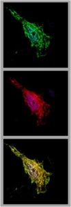
Colocalisation of MyrB (red) with
GFP-tagged NTCP (green) in HepG2 cells
Project Staff

Benno Zehnder, PhD

Florian Lempp, PhD