HIV-1 capture and spread at nanoscopic resolution

Project description – Summary – 3rd funding period
We apply advanced microscopic techniques to characterize critical steps in early HIV-1 replication on the single particle level with high spatial resolution in physiologically relevant cell systems.
We have shown that apparently intact capsids enter the nucleus through intact nuclear pores in all cell types analyzed, and uncoating of the viral genome occurs by rupture of the capsid inside the nucleus. Building on these observations, which have changed the general view of early HIV-1 replication, we will interrogate the hypothesis that intact capsids orchestrate the entire early phase of HIV-1 replication. Furthermore, we will investigate the process of genome uncoating in the nucleus by rupture of the capsid lattice.
Wir verwenden moderne Mikroskopie mit höchster räumlicher Auflösung zur Charakterisierung der frühen HIV-1 Replikation auf Einzelpartikel-Ebene in relevanten Zellsystemen.
Wir haben gezeigt, dass anscheinend vollständige Kapside den Zellkern durch intakte Kernporen erreichen und Uncoating des viralen Genoms durch Aufbrechen des Kapsids im Nukleus geschieht. Auf der Grundlage dieser Ergebnisse, die das Verständnis der frühen HIV-1 Replikation grundsätzlich verändert haben, werden wir jetzt die Hypothese überprüfen, dass das intakte Kapsid alle Schritte der frühen Replikation organisiert. Des Weiteren werden wir den Prozess und Mechanismus des Genom-Uncoating im Zellkern untersuchen.
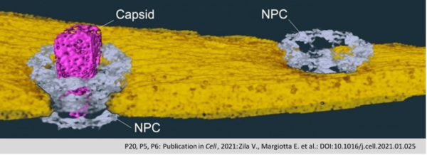
The protein capsule of the HIV-1 (Capsid; pink) passes as a whole through the nuclear pore
(NPC; grey) into the cell nucleus, where it disintegrates and releases the viral genetic material.
The nuclear membrane is shown in yellow. (Zila et al., Cell 2021).
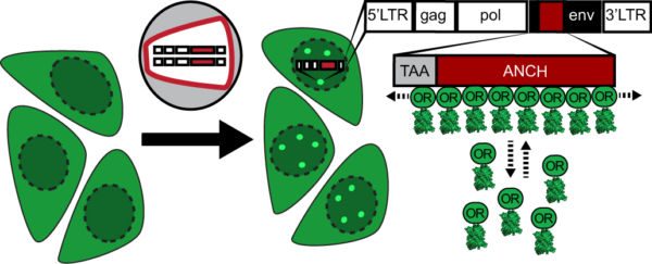
Scheme of the ANCHOR dsDNA visualization system. Fluorescently tagged OR3 binds to the ANCH sequence introduced into the HIV-1 genome.

Imaging of HIV-1 cDNA signal formation in living cells

CLEM-ET renderings of conical and capsid-like HIV-1 complexes inside the nucleus. Internal electron-density indicates the presence of the RNP complex in apparently intact capsids. Tubular and ruptured capsids often appear empty (right panel, bottom capsid). The middle panel shows a positions that correlated to both, fluorescently tagged IN and the OR3 marker, and might show a capsid captured in the process of uncoating.
Tomographic reconstruction and isosurface rendering highlighting a conical capsid within a nuclear pore complex. capsid, magenta; nuclear envelope, yellow; NPC, cyan.
.
Project staff
Lara Rohleder
PhD student
Jian Song
PhD student
Samara Martin Alonso
Postdoc