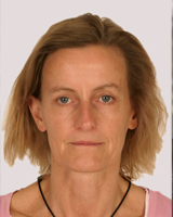
Priv.-Doz. Dr. Jacomine Krijnse-Locker
Jacomine Krijnse-Locker has led Project Z2 together with John Briggs from October 2014-April 2015.
Since May 2015 her group is located at the Institut Pasteur, Paris.
until April 2015
Electron microscopy core facility and Department of Infectious Diseases, Virology
Heidelberg University
69120 Heidelberg, Germany
FIELDS OF INTEREST
Virology, assembly of large DNA viruses, cell biology, dynamics of membranes, cell biology of viruses, electron microscopy, imaging of viral and cellular membranes, 3D microscopy
AWARDS & HONORS
1990-1994 | PhD fellowship ‘Dutch organization for scientific research’ (NWO) |
1994-1996 | Post-doctoral fellowship from ‘Human science frontiers program organization’ |
2015-present | Head of the unit for technology and service in ultra-structural bio-imaging, Institut Pasteur, Paris |
2009-present | co-head of the EM core facility of Heidelberg University |
2007-present | Group leader at the Department of Infectious Diseases, Virology &Molecular Virology, Heidelberg University |
1997-2006 | Scientist, EMBL, Heidelberg |
2003 | Habilitation, Heidelberg University (cell biology) |
1994-1997 | post-doctoral fellow EMBL, Heidelberg, cell biology program |
1990-1994 | PhD in virology and cell biology, University of Utrecht, Utrecht, theNetherlands |
2001 | Main organizer of EMBO-workshops ‘Cell Biology of Virus Infections’, EMBL, Heidelberg, September 2001 |
2004-2007 | co-head (together with Dr. Stephan Urban) of the study section cellbiology of the German society for Virology |
2004 | Main organizer second EMBO workshop “Cell Biology of Virus Infections’. EMBL, Heidelberg, September 2004 |
Shah PNM, Stanifer ML, Hohn K, Engel U, Haselmann U, Bartenschlager R, Kräusslich HG, Krijnse-Locker J, Boulant S (2017). Genome packaging of reovirus is mediated by the scaffolding property of the microtubule network. Cellular Microbiology doi: 10.1111/cmi.12765. [Epub ahead of print]
Chlanda P, Krijnse Locker J (2017). The sleeping beauty kissed awake; new methods in electron microscopy to study cellular membranes. Biochemical J. 474(6):1041-1053
Chlanda P, Carbajal MA, Cyrklaff M, Griffiths G, Krijnse Locker J (2009). Membrane rupture generates single open membrane sheets during vaccinia virus assembly. Cell Host & Microbe 6, 81-90
Pan X, Rudolph JM, Abraham L, Habermann A, Haller C, Krijnse-Locker J, Fackler OT (2012). HIV-1 Nef compensates for disorganization of the immunological synapse by inducing trans-Golgi network-associated Lck signaling. Blood 119, 786-797.
Romero-Brey I, Merz A, Chiramel A, Lee JY, Chlanda P, Haselman U, Santarella-Mellwig R, Habermann A, Hoppe S, Kallis S, Walther P, Krijnse Locker J, Bartenschlager R (2012). Three-dimensional architecture and biogenesis of membrane structures associated with hepatitis C virus replication. PLoS Pathog 8, e1003056.
Suárez C, Welsch S, Chlanda P, Hagen W, Hoppe S, Kolovou A, Pagnier I, Raoult D, Krijnse Locker J (2013). Open membrane-intermediates are the precursors for assembly of large DNA viruses. Cell Microbiol. 15(11) :1883-95. doi: 10.1111/cmi.12156. Epub 2013 Jul 5.
Welsch S, Miller S, Romero-Brey I, Merz A, Bleck CKE, Walther P, Fuller SD, Antony C, Krijnse-Locker J, Bartenschlager R (2009). Composition and three-dimensional architecture of the dengue virus replication and assembly sites. Cell Host & Microbe 5, 365-375
Welsch S, Keppler OT, Habermann A, Allespach I, Krijnse Locker J, Kräusslich HG (2007). The primary site of HIV-1 budding in infected primary macrophages is the plasma membrane. PLOS Pathog 3, 1-11
Click here for further publications.